Gino Fornaciari*, Lorenzo Costantini**, Rosalba Ciranni*
*Department of Oncology, Transplants and Advanced Technologies in Medicine
Section of History of Medicine and Paleopathology, University of Pisa, Via Roma 57, 56126 Pisa, Italy
**National Museum of Oriental Art, Laboratory of Paleobiology, Via Merulana 248, 00185 Roma, Italy
Keywords: trauma, bullet-hole, dressing, natural mummy
ABSTRACT
The crypt of the church of the "Saints Jesus and Mary" of Borgo Cerreto, a village near Spoleto (Umbria, central Italy), revealed the natural mummy of a soldier, dated at the first of the 19th century. The inferior half of the right thigh showed a large circular dressing. X-ray evidenced a comminuted fracture of the inferior third of the right femoral diaphysis, with the presence of small metallic fragments. A medication still in situ was present under the dressing. The wound of oval shape (2 x 1.6 cm in size), the comminuted fracture of the diaphysis and the tiny metallic fragments prove that we are in the presence of a gunshot wound. The size of the wound, well adapting to the 16 mm gauge of the military gun balls of the first half of the 19th century, allowed us to identify it with a typical bullet-hole.
CASE REPORT
The crypt of the church of the "Saints Jesus and Mary" of Borgo Cerreto, a village near Spoleto (Umbria, central Italy), revealed six natural mummies in good state of preservation, dating back to the 18th and at the first of 19th century.
One of the bodies belonged to a man, buried in stretched out position, slightly curved to the left and with the inferior limbs bent. The individual in question was a man of medium-high stature (about 170 cm) (Trotter and Gleser, 1958), who died between 25 and 35 years of age (Fig. 1 a,b). The style of the garment, consisting in a long jacket, tightened around the waist, a shirt with very large sleeves and trousers with buttons, worn underneath the boots, made it possible to date the mummy, most probably a soldier, at the end of the 18th century or at the first of the 19th century (Fig. 1 c).

Fig. 1 The mummy at the moment of discovery (a); a graphic reconstruction of the body (b) and the possible aspect of the individual in life (c).
The right wrist presented a dressing, covering a transversal wound with enlarged margins. X-ray, performed in loco with a portable radiographic apparatus, did not evidence any lesion of the bones of the right forearm and arm; the total absence of vertebral arthritis confirmed that the subject was still young.
THE WOUND
The inferior half of the right thigh revealed a large circular dressing, fastened with many small nodes (Fig. 3a). X-ray evidenced a comminuted fracture limited to the inferior third of the femoral diaphysis, with the presence of small metallic fragments (Fig. 2). A medication still in situ was present under the dressing, with a plug penetrating through the cutaneous muscular layers (Fig. 3b).
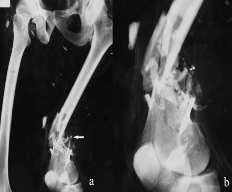
Fig. 2 X-ray of the right thigh with a comminuted fracture of the inferior third of the femoral diaphysis and small metallic fragments (arrow).
After plug removal, a wound appeared of the lateral region of the thigh. The wound was of oval shape, 2 x 1.6 cm in size, revealing the bone splinters of the femoral diaphysis, already evidenced by X-ray (Fig. 3 c).
The opening of the cutaneous and muscular layers of the thigh confirmed that these were the bone splinters of the femoral diaphysis, as already evidenced by X-ray.

Fig. 3 Large circular dressing of inferior half of the right thigh, fastened with many small nodes (a), with a plug penetrating through the cutaneous muscular layers (b, arrow) and an oval wound (2 x 1.6 cm); the bone splinters of the femoral diaphysis are evident (c, arrow).
Even the inferior half of the left thigh, completely skeletonized, presented a circular dressing with a plug, without any lesions of the femur.
CONCLUSIONS
The oval lesion of the thigh, the comminuted and circumscribed fracture of the diaphysis of the right femur and, in particular, the tiny metallic fragments, proves that we are in the presence of a gunshot wound (Spitz, 1993). The circular shape and the size of the wound, well adapting to the 16 mm gauge of the military gun balls of the second half of the 18th century (Exteberria, 1999; Lunardini et al., 2002), allowed us to identify it with a typical bullet-hole.
Therefore, during a combat, the individual was seriously injured by a bullet which penetrated laterally, causing the exposed and comminuted fracture of the femur. Even the lesion on the right pulse is almost certainly referable to the same event.
Despite the professional cures, the injured person died, probably of complications.
The authors acknowledge Dr. Roberta Faggioni for figures, Mr. Fabio Cini for graphic reconstructions and Mr. Marcello Gambini for technical assistance.
LITERATURE CITED
Berryman HE, and Simes SA. 1998. Recognition of gunshot and blunt cranial trauma through fracture interpretation. In: Reichs KJ, editor. Forensic Osteology. Advances in the identification of human remains. Springfield, Illinois: Thomas Publisher, p. 333-352.
Exteberria F. 1999. Surgery in the Spanish war independence (1807-1813) between Desault and Lister. J Paleopath 11: 25-40.
Lunardini A, Rollo F and Fornaciari G. 2002. Bone lesions from the ossuary of the Napoleonic battle of Marengo, Northern Italy (14th June 1800). J Paleopath 14: 69-75.
Spitz WE. 1993. Injury by gunfire. In: WU Spitz Ed., Medicolegal investigation of death. Springfield, Illinois: Thomas Publisher, p. 311-412.
Trotter M, and Gleser GC. A re-evaluation of estimation of stature based on measurements of stature taken during life and of long bones after death. Am J Phys Anthropol 16: 79-123.


